
Stress Tests
Stress tests are performed to determine how your heart reacts to working harder (the stress portion). Stress tests can be performed for a variety of reasons but the most common reason is to determine if there’s adequate blood supply going to the heart muscle. As you exercise or perform exertional activities, your body requires more fuel (oxygen brought by blood) and your heart has to pump more blood. If the arteries that supply the heart have blockages, the heart will not get enough blood and patients will develop EKG changes, chest pain, or abnormal imaging.
There are several different types of stress tests, and not all require you to walk on a treadmill.

Treadmill Stress Test
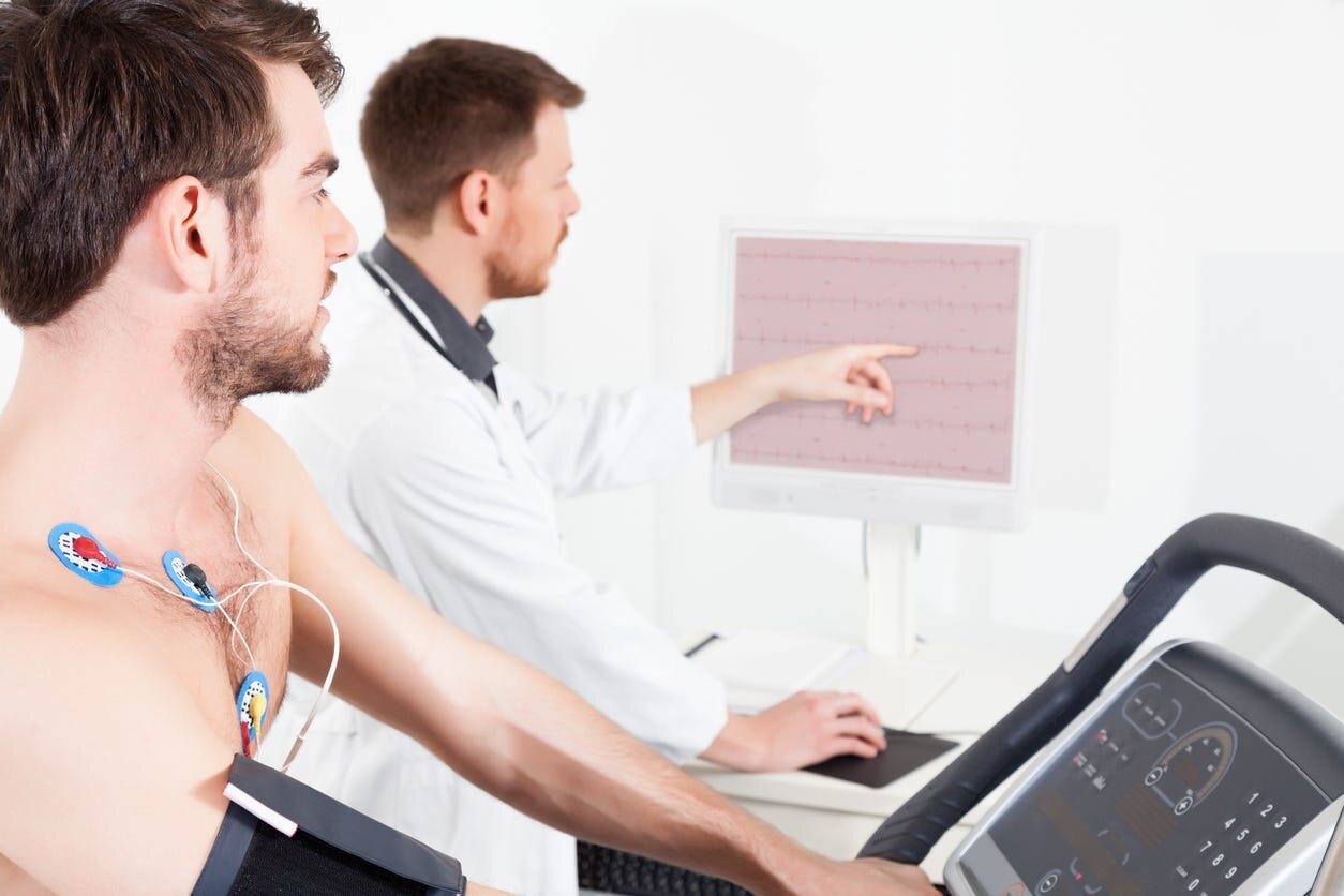
Stress tests can be performed in a variety of different ways. The most basic stress test is a Treadmill stress test. A patient is hooked up to EKG electrodes as well as a blood pressure (BP) cuff. Recordings of both vitals and EKG are obtained as you progress during the stress test. The treadmill starts at a slow place, but every three (3) minutes it will get gradually higher and faster. You can walk as long as you can tolerate but a minimum target to achieve is a pre-specified target heart rate (THR) that will be calculated based on your age. If you are unable to achieve this target or feel any discomfort, the test can be stopped early.
What to Wear:
Wear comfortable “gym” clothing and walking or tennis shoes with rubber soles.
Do not drink alcohol or caffeine before this or any type of stress test.
You may be asked to temporarily hold certain medications (beta-blockers: metoprolol, carvedilol) but do not stop before checking with your doctor.

Exercise Stress Echocardiogram
An exercise stress echocardiogram (ESE) is very similar to a basic treadmill however ultrasound imaging of your heart is obtained before and after the stress portion of the treadmill.
Steps of an ESE
The test is performed by the patient taking a baseline ECG (EKG) as well as resting vital signs (BP and HR).
Ultrasound images of your heart are obtained by the patient laying on their left side. The doctor is looking for how the walls of the heart are moving and pumping at rest.
The patient then walks on the treadmill using the same protocol as above until the THR is achieved (or as long as the patient can go).
Once the target is achieved, patient returns to the table and a second set of images are obtained. Again, the same views of the heart are obtained to see how the walls of the heart are moving.
A normal stress echo test would show the heart pumping stronger and faster after exercise. If there is a problem such as a coronary blockage that portion of the heart muscle may pump weaker than it should. This can also occur if this part of the heart previously had a heart attack.

Chemical Nuclear Stress Test (aka Lexiscan Stress Test)
The final type of stress test is usually reserved for patients who are unable to walk on the treadmill or require special imaging due to their prior history of heart disease. This type of stress test, can use a chemical agent to “stress” the heart, and replicate when a patient is unable to walk and bring up the heart rate naturally. The most common agent given is Lexiscan. However, patients can also walk on the treadmill instead of using the Lexiscan medication if feasible. A very small dose of a radioactive tracer is also given to “light” up the heart.
Steps of a Lexiscan Chemical Nuclear Stress Tests
1. The test is performed by the patient taking a baseline ECG (EKG) as well as resting vital signs (BP and HR).
2. The patient is injected with a small dose of a radioactive dye, and images of the heart are obtained.
3. Then Lexiscan (the stressing agent) is administered to the patient. Alternatively the patient may walk on the treadmill if they are able to reach their Target Heart Rate.
4. At peak exercise another small dose of the radioactive dye is given.
5. The patient is again imaged with the nuclear camera during this “stress’ portion.
6. The differences in imaging following the rest portion and the stress portion are